The center of your vision starts to look a little blurry. Straight lines on doorframes appear slightly wavy. Reading becomes harder not because the words are smaller but because something sits in the middle of your view making them hard to focus on. Faces look normal until you try to recognize fine details, and then something feels off.
Age related macular degeneration, usually called AMD or just macular degeneration, is the leading cause of vision loss in adults over fifty in developed countries. It does not cause complete blindness in most cases, but it damages the central vision that matters most for reading, driving, recognizing faces, and daily tasks. Understanding how it works, who gets it, and what slows or halts its progression can meaningfully change the trajectory of your vision as you age.
The Macula and Why It Matters
The macula is the central region of the retina, the light sensing tissue at the back of the eye. Though only a few millimeters across, the macula handles all the sharp central vision you use for reading, detailed work, and recognizing what you are looking at directly. The peripheral retina provides the broader field of vision that gives you awareness of your surroundings.
When the macula is damaged, central vision suffers while peripheral vision remains intact. This creates a distinctive pattern where people can still walk around and see the general shape of things but cannot read fine print, see fine details at a distance, or recognize faces clearly. The world has a blurry spot in the middle that they have to look around.
The Two Main Types
AMD comes in two primary forms. Dry AMD, also called atrophic AMD, is the more common type, accounting for roughly eighty five percent of cases. It progresses slowly over years, typically moving through early, intermediate, and late stages. In late dry AMD, areas of the macula atrophy and the photoreceptors die.
Wet AMD, or neovascular AMD, is less common but more aggressive. Abnormal blood vessels grow beneath the retina and leak fluid or blood, damaging the macula rapidly. Without treatment, wet AMD can cause severe central vision loss within weeks to months. With treatment, outcomes have improved dramatically in recent years.
Some people start with dry AMD and later develop wet AMD. Monitoring is important because catching the wet form early matters for preserving vision.
Early Warning Signs
AMD often starts silently. Early stages may produce no symptoms at all, detected only on routine eye exams by changes visible in the macula called drusen, small yellow deposits beneath the retina.
As AMD progresses, symptoms begin. You might notice that straight lines appear wavy or bent, a finding called metamorphopsia. This is a hallmark of AMD affecting the macula. The edge of a door, a telephone pole, or text lines in a book look crooked when they should be straight.
A blurry or blank spot may appear in the center of vision. At first subtle, it gradually enlarges or darkens. Reading becomes effortful because the text in the very center is distorted or missing.
Colors may seem less vibrant than they used to. Contrast perception fades. Faces appear increasingly hard to recognize at conversational distance.
Difficulty adapting to low light is common. Moving from a bright outdoor setting to a dim restaurant becomes disorienting. Reading in anything less than strong light becomes challenging.
Many AMD patients compensate without realizing it, using peripheral vision more, turning their head to view things with a better part of the macula, avoiding reading because it is tiring. They may not mention symptoms until advanced stages.
A home test called the Amsler grid helps detect changes. It is a simple grid pattern you look at with one eye at a time while focusing on a central dot. Any waviness, missing squares, or distortion suggests macular changes that warrant prompt evaluation.
Risk Factors
Age is the biggest risk factor. AMD becomes increasingly common after fifty and rises sharply after seventy five. Most cases are clearly age related, though earlier onset does occur in some families.
Genetics play a huge role. Having a first degree relative with AMD significantly increases your risk. Certain gene variants, particularly in the complement factor H gene, strongly predispose to AMD. Genetic testing is available and sometimes informative, though interpretation is complex.
Smoking is the strongest modifiable risk factor. Smokers develop AMD earlier and progress faster. The damage appears to be cumulative, so stopping at any age reduces ongoing risk. Secondhand smoke matters too.
Diet influences risk substantially. Diets low in antioxidants, omega three fatty acids, and green leafy vegetables correlate with higher AMD rates. The Mediterranean dietary pattern is associated with lower risk.
Obesity and cardiovascular disease both elevate AMD risk, likely through shared vascular mechanisms. High blood pressure and high cholesterol matter.
Ultraviolet and blue light exposure may contribute, though evidence is less conclusive than for other risk factors. Regular use of quality sunglasses makes sense given the small cost.
Lighter iris color and European ancestry are associated with higher AMD rates. This is not something you can change, but it reinforces the importance of other preventive measures if you fit this profile.
Getting Properly Screened
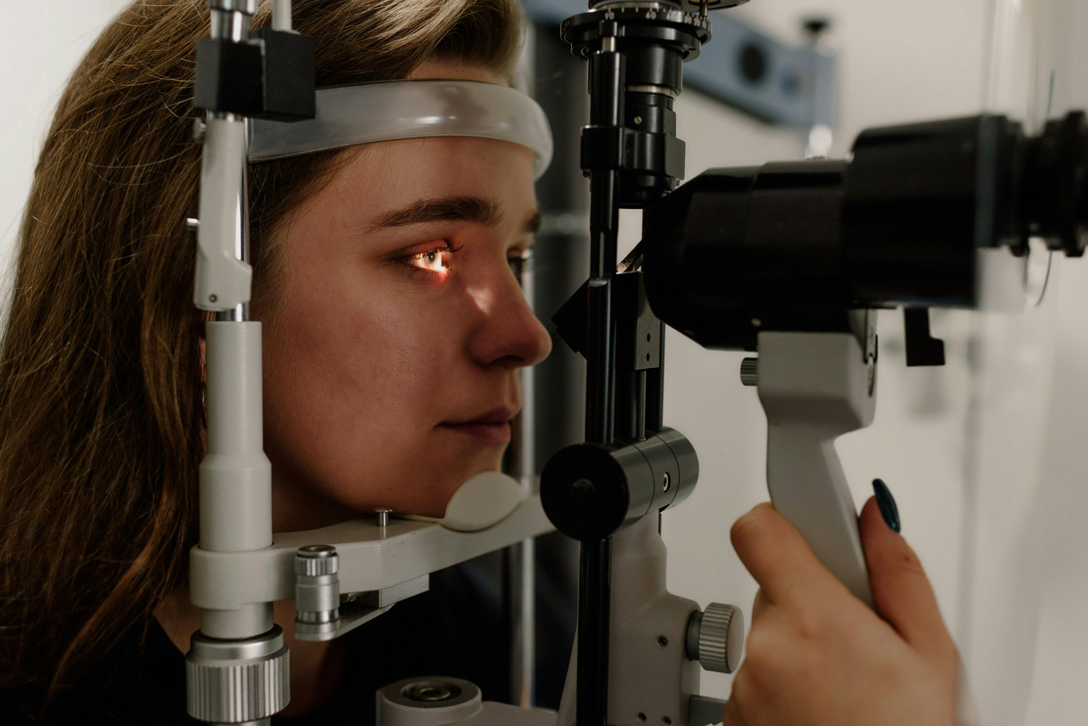
AMD screening is part of comprehensive eye exams. Your eye doctor dilates your pupils and examines the macula with specialized instruments. Early changes like drusen are visible before symptoms appear.
Optical coherence tomography, or OCT, has transformed AMD monitoring. This painless scan provides cross sectional images of the retina, showing drusen size, macular thickness, and signs of fluid accumulation that indicate wet AMD. OCT has become standard for AMD evaluation and monitoring.
Fundus autofluorescence imaging detects areas of unhealthy retina before structural damage is obvious. Fluorescein angiography, which involves injecting a dye and photographing the retina, is used when wet AMD is suspected to map abnormal blood vessels.
Risk assessment takes all this information together. Large drusen, pigment changes, and family history all factor into predicting progression. Your doctor can estimate your chance of advancing to late AMD over the next few years, which guides monitoring intensity and treatment decisions.
Preventing Progression
Once AMD is diagnosed, the focus shifts to slowing progression. Several interventions have strong evidence.
The AREDS2 supplement formulation has the best evidence for intermediate to advanced dry AMD. It contains vitamin C, vitamin E, lutein, zeaxanthin, zinc, and copper in specific amounts. Trials showed it reduced progression to late AMD by about twenty five percent in appropriate candidates. It does not prevent AMD in people without existing disease, and beta carotene containing older formulations should be avoided in smokers due to lung cancer risk.
Smoking cessation is essential. The benefit begins immediately even for long term smokers. Any progress toward quitting helps.
Dietary patterns rich in dark leafy greens, colorful vegetables, fatty fish, nuts, and olive oil reduce progression. Eat the rainbow daily. Salmon, sardines, and mackerel provide omega three fatty acids that support retinal health.
Blood pressure and cholesterol management matter for general cardiovascular health and may slow AMD progression specifically.
Exercise regularly. Physical activity is associated with lower AMD rates and slower progression, possibly through improved circulation and inflammation control.
Protect your eyes from intense sunlight. Quality sunglasses with UV protection and blue light reduction, worn consistently outdoors, are low cost insurance.
Monitor yourself at home with an Amsler grid. Check each eye separately, focusing on the central dot, and note any new distortion. Sudden changes warrant same week or same day eye doctor evaluation to rule out wet AMD.
Treatment Options
For dry AMD, specific treatment options have been limited until recently. Supplementation and lifestyle remain the foundation. New treatments for geographic atrophy, the late stage of dry AMD, have been approved. Pegcetacoplan and avacincaptad pegol injections slow geographic atrophy progression, though they do not improve vision and involve monthly or bimonthly injections for years. These treatments are new and decisions about starting them require nuanced conversation with a retina specialist.
For wet AMD, treatment has been transformed by anti VEGF injections. These medications, including aflibercept, ranibizumab, bevacizumab, and newer agents like faricimab, shut down the abnormal blood vessels that cause damage. Treatment is given by injection directly into the eye, typically monthly or every few months depending on response and medication chosen. The injections are generally well tolerated despite sounding uncomfortable.
Results from anti VEGF treatment vary. Many patients gain back vision that was lost in the initial wet AMD episode. Others stabilize at the level they were at when treatment began. A minority continue to lose vision despite treatment. Early intervention produces the best results, reinforcing why prompt evaluation of sudden changes matters.
Laser treatments and photodynamic therapy are used rarely for specific situations where anti VEGF alone is inadequate.
Living With Vision Loss
For those with advanced AMD and permanent central vision loss, low vision rehabilitation makes an enormous difference. Specialized optometrists and therapists help maximize remaining vision through tools like magnifiers, stronger lighting, high contrast aids, and training in eccentric viewing techniques that use peripheral vision more effectively.
Technology continues to improve. E readers with adjustable text size, voice control for devices, audiobooks, and specialized apps all help maintain independence. Speaking watches, audio scales, and talking glucometers are available for daily tasks.
Many communities have low vision support groups. Sharing experiences with others who understand helps reduce the isolation that vision loss can bring.
Emotional adjustment matters. Grief over lost abilities is normal. Depression is common and treatable. Counseling focused on adaptation can help significantly.
Looking Ahead
Research on AMD is active and promising. New drugs are in development. Stem cell therapies are being studied. Gene therapies for specific genetic forms of AMD show early promise. Retinal prostheses and brain interfaces are beginning to offer limited restoration of vision in the most severely affected.
For now, the combination of smoking cessation, quality nutrition, appropriate supplementation when indicated, prompt treatment of wet AMD, and regular monitoring represents the best approach. Most people with AMD can preserve useful vision for many years with proper care, even as the disease slowly progresses.
The Bottom Line
Macular degeneration is common, often quietly progressive, and responsive to both prevention and treatment strategies. If you have family history, if you smoke, if you are over fifty and have not had a comprehensive eye exam recently, do not wait for symptoms. Early detection and appropriate intervention protect the central vision that makes so much of life possible. Your vision is worth the effort.
Sources and Further Reading
Health and Beyond uses reputable medical and scientific sources where possible. These links support or expand on the topics discussed above.
- National Eye Institute: Eye Healthnei.nih.gov
- MedlinePlus: Eyes and Visionmedlineplus.gov