Your vision keeps changing. Every year you need a new glasses prescription, and even with the new lenses, things never look quite right. Contact lenses do not feel right either. Lights have multiple ghostly images trailing off them at night. Your eye doctor mentions something called keratoconus, and suddenly a condition you have never heard of becomes part of your life.
Keratoconus is a progressive eye condition where the normally round cornea thins and bulges into a cone shape. This distortion scrambles the light entering the eye and causes vision problems that conventional glasses cannot fully fix. It affects roughly one in two thousand people, typically starting in the teenage years or early twenties and progressing over years to decades. The outlook for keratoconus patients has improved dramatically in recent years thanks to new treatments that can halt progression and restore vision.
What Is Actually Happening
The cornea is the clear dome at the front of the eye that does most of the eye focusing work. A healthy cornea has a regular curvature that bends light cleanly onto the retina. In keratoconus, the cornea progressively thins, particularly in a localized area, and bulges outward into a cone shape. This irregularity distorts the light entering the eye and causes irregular astigmatism, a complex optical error that glasses struggle to correct.
The thinning appears to involve abnormalities in the proteins that provide corneal strength, particularly collagen. Genetic, environmental, and mechanical factors all contribute. Eye rubbing is now recognized as a significant factor in progression, and anyone with known or suspected keratoconus should avoid rubbing their eyes.
Keratoconus is bilateral, meaning it affects both eyes, but often asymmetric. One eye may be significantly worse than the other, and progression timing may differ between eyes.
Who Gets It
Keratoconus usually begins in the teens or twenties. Progression typically continues for a decade or two before often stabilizing, though this varies widely. Earlier onset tends to mean more aggressive progression.
Family history increases risk. Having a close relative with keratoconus raises your risk ten fold or more. Several genes have been associated, and genetic research continues.
Eye rubbing is a major modifiable risk factor. Chronic hard eye rubbing, often driven by allergies or dry eye, contributes to keratoconus development and progression. Breaking the rubbing habit is crucial for anyone diagnosed.
Allergies and atopic disease, including asthma, eczema, and hay fever, are strongly associated with keratoconus. The connection may involve eye rubbing from itch plus underlying inflammatory tendencies.
Down syndrome carries a significantly higher risk of keratoconus.
Connective tissue disorders like Marfan syndrome, Ehlers Danlos syndrome, and osteogenesis imperfecta have higher keratoconus rates.
Sleep apnea has been associated with keratoconus, possibly through eye rubbing during partial awakenings or through inflammatory mechanisms.
The Early Symptoms
Early keratoconus is often subtle. Your vision quality deteriorates gradually despite apparently adequate correction. Glasses prescriptions change frequently. Contact lenses that used to be comfortable become difficult to fit.
Streaks, ghosting, or multiple images around lights, especially at night, are classic early symptoms. Streetlights and headlights appear to have fingers extending from them. This is particularly noticeable when driving at night.
Sensitivity to bright light increases. Reading becomes effortful. Eye strain after visual tasks develops.
The combination of normal or mildly blurry vision on routine testing with complaints of visual quality problems should raise suspicion of keratoconus. Standard eye exams using a letter chart may not reveal the full picture. Corneal topography, which maps the corneal surface curvature, is the key diagnostic test.
More advanced keratoconus produces obviously distorted vision that significant glasses prescriptions cannot fully correct. Some patients develop corneal hydrops, where sudden swelling of the cornea causes acute vision blur, pain, and a white spot visible in the cornea. This resolves slowly but leaves scarring.
Getting Properly Diagnosed
Corneal topography is the foundation of keratoconus diagnosis. This painless test creates a detailed map of your corneal curvature, showing the characteristic pattern of keratoconus and quantifying its severity. It can detect early keratoconus before symptoms are severe.
Corneal tomography using Scheimpflug imaging or anterior segment OCT provides additional detail about corneal thickness across the entire surface, not just the curvature. Progressive thinning in a specific location is a hallmark of keratoconus.
Refraction, the process of determining your best glasses prescription, reveals the irregular astigmatism typical of keratoconus. Vision that does not correct to normal even with best glasses is another clue.
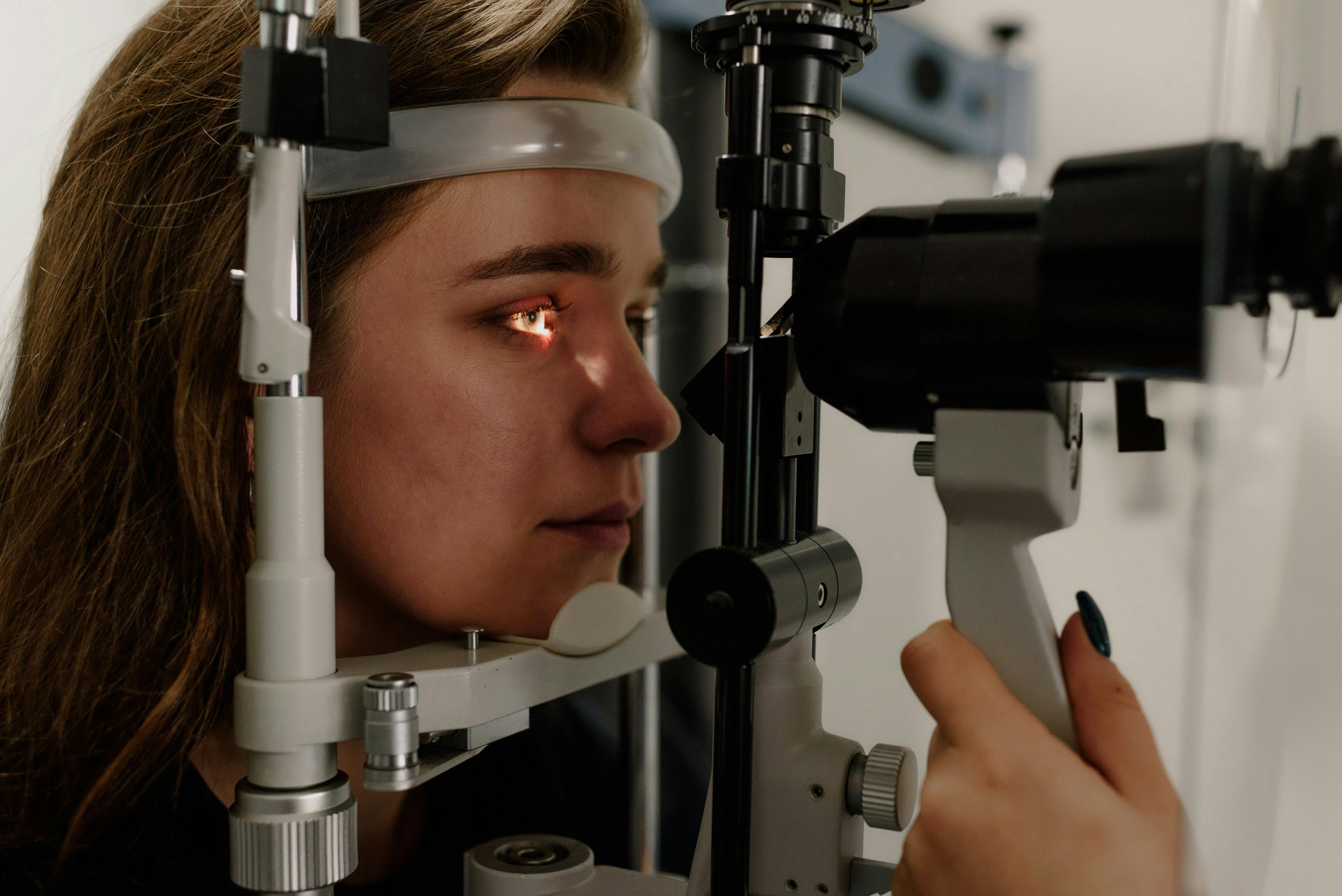
Slit lamp examination by an ophthalmologist can reveal specific findings like Vogt striae, stress lines in the cornea, Fleischer rings of iron deposition, and the corneal thinning itself in more advanced cases.
Specialized contact lens fitting may be part of evaluation since standard lenses often do not fit well.
Halting Progression
The treatment landscape for keratoconus has been transformed by corneal cross linking, a procedure that strengthens the cornea and halts progression in most patients. Cross linking uses riboflavin drops and UV light to create additional bonds between collagen fibers in the cornea, essentially stiffening the tissue.
Cross linking is most valuable for patients with documented progression, typically younger patients in their teens, twenties, or thirties. Stopping progression before significant visual damage occurs preserves options and protects vision.
The procedure takes about an hour, with a recovery period of a few days of significant discomfort and several weeks of healing. Results are generally excellent, with stabilization of the cornea in the great majority of treated eyes. Vision may improve slightly in some patients, though cross linking is primarily about stopping progression rather than restoring vision.
Variations of the cross linking protocol have emerged, with different UV intensities, durations, and epithelium handling approaches. Your surgeon tailors the approach to your situation.
Alongside cross linking, avoiding eye rubbing is essential. Treat underlying allergies, dry eye, and other causes of eye irritation to reduce the urge to rub. If you rub, train yourself out of the habit through awareness and alternatives.
Correcting Vision With Keratoconus
In early keratoconus, glasses may provide adequate vision. As the cornea becomes more irregular, glasses become less effective because they cannot correct the irregular astigmatism.
Soft contact lenses work for mild keratoconus sometimes but usually fail to provide good vision as the condition progresses.
Rigid gas permeable, or RGP, contact lenses have been a mainstay for decades. The rigid lens creates a smooth optical surface over the irregular cornea, providing much better vision than glasses or soft lenses. Getting a good fit takes skill and sometimes many adjustments, but well fitted RGPs transform vision for many keratoconus patients.
Hybrid lenses combine an RGP center with a soft skirt, offering some advantages for comfort and stability.
Scleral lenses have revolutionized keratoconus contact lens fitting. These large diameter lenses vault entirely over the cornea and rest on the white of the eye. They do not touch the sensitive cornea at all, providing exceptional comfort even in severe disease. The space between lens and cornea is filled with saline, creating a liquid tear layer that masks all corneal irregularity. Vision through a well fit scleral lens is often dramatically better than anything else can provide.
Scleral lens fitting requires specialized training. Finding an experienced fitter is worth the effort. Many keratoconus patients who struggled with other lens types thrive in sclerals.
Surgical Options for Advanced Disease
For severe keratoconus that does not respond adequately to contact lenses, several surgical options exist.
Intracorneal ring segments, small plastic rings implanted into the cornea, can flatten and regularize the corneal shape. They reduce irregular astigmatism and improve contact lens fitting in some patients.
Phakic intraocular lenses implanted inside the eye can correct high refractive errors in stable keratoconus.
Corneal transplantation was historically the standard for severe disease. Full thickness penetrating keratoplasty replaces the entire cornea with a donor. Partial thickness procedures like deep anterior lamellar keratoplasty, DALK, replace only the diseased layers while preserving the inner cornea, reducing risks of rejection and faster visual recovery.
Modern transplant techniques have excellent outcomes, but recovery takes many months, and lifelong eye drops may be needed. Transplants can also develop their own problems over time. These procedures are reserved for cases where less invasive options are inadequate.
Living With Keratoconus
Regular eye care is essential. Ongoing monitoring catches progression early when cross linking can help most. Your eye doctor should document corneal topography periodically and watch for changes.
Cross linking once done is typically durable, but occasionally progression continues and retreatment is considered.
Contact lens adjustments may be needed periodically as the cornea changes. A good lens fitter becomes a long term partner in care.
Dry eye and allergies need active management to protect the eyes and reduce rubbing urges.
Sun protection with quality UV blocking sunglasses is worthwhile, as UV exposure may influence corneal health.
Emotional adjustment matters. Dealing with progressive vision problems starting in young adulthood can be stressful. Support groups and online communities help reduce isolation.
Questions About Children
Early onset keratoconus in children and teens tends to progress faster. Lower threshold for cross linking is reasonable in pediatric patients because progression risk is higher and long term visual preservation matters especially for young eyes.
Close monitoring with frequent topography catches progression early in this age group.
Avoiding eye rubbing is particularly important in children who may rub more automatically.
When to See a Specialist
If you have been diagnosed with keratoconus, getting care from a cornea specialist is reasonable. These ophthalmologists have specific training and expertise beyond general eye care.
If you have keratoconus symptoms and your current provider has not done corneal topography or cannot interpret it, seek a second opinion.
If your vision is deteriorating despite current treatment, ask about cross linking if you have not had it, sclerals if you have not tried them, or surgical options for advanced cases.
Looking Ahead
Keratoconus research continues to advance. Better imaging allows earlier detection. Refinement of cross linking protocols improves outcomes. New contact lens designs expand options. Gene therapies are being studied for patients with specific genetic causes. Regenerative approaches to rebuild corneal tissue are in development.
The outlook for keratoconus today is far better than it was even a decade ago. Most patients, especially those diagnosed early and treated appropriately, maintain good functional vision throughout life.
The Bottom Line
Keratoconus is a serious condition but a manageable one with modern care. Early diagnosis allows cross linking to preserve the cornea. Scleral lenses transform vision for most patients. Surgery is rarely needed and produces good results when it is. If you suspect keratoconus, seek specialized evaluation. Protecting your vision at this stage has long lasting benefits. The cornea can be stabilized and your sight preserved.
Sources and Further Reading
Health and Beyond uses reputable medical and scientific sources where possible. These links support or expand on the topics discussed above.
- National Eye Institute: Eye Healthnei.nih.gov
- MedlinePlus: Eyes and Visionmedlineplus.gov