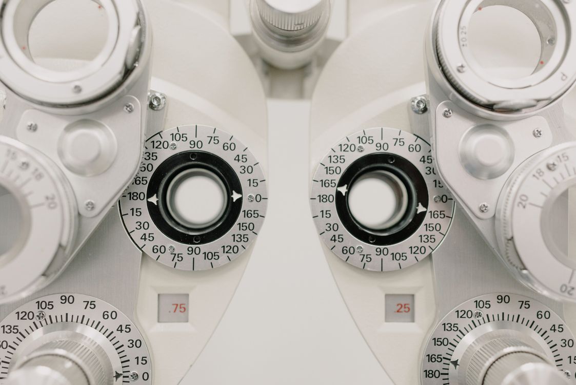
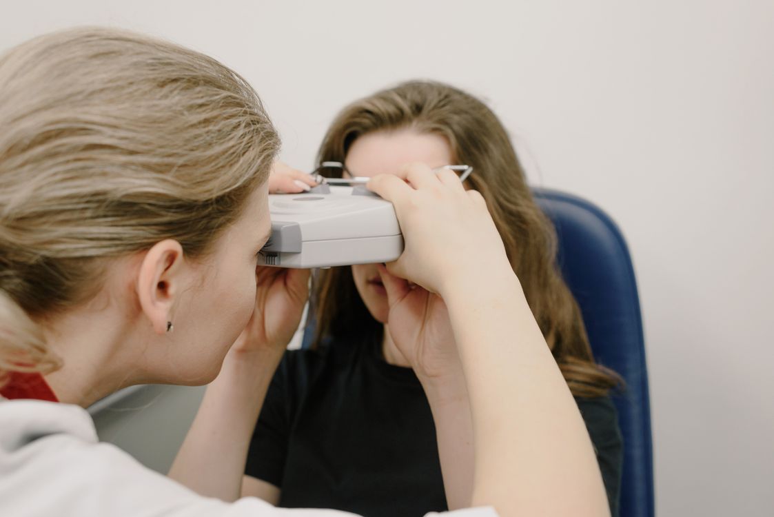
If you live with diabetes, your eyes are working overtime. Every time blood sugar climbs, the delicate web of blood vessels at the back of your eye takes a hit. Most people never feel it happening. That silence is exactly what makes diabetic eye disease so dangerous, and it is also why it has quietly become the number one cause of preventable blindness in working-age adults across the United States and much of the world.
The good news is that vision loss from diabetes is not inevitable. When it is caught early, the vast majority of serious damage can be stopped or reversed. The trick is knowing what diabetic eye disease actually is, which exams pick it up, how often to get screened, and which daily habits keep your retina healthy for decades.
This guide walks you through all of that in plain language, with the numbers and timelines your eye doctor wishes every patient already knew.
What Diabetic Eye Disease Actually Means
Diabetic eye disease is an umbrella term. It is not one condition. Under that umbrella sit four separate problems, and a person with diabetes can develop any combination of them.
Diabetic retinopathy is the most common and the most feared. High blood sugar damages the tiny blood vessels that feed the retina, the light-sensing tissue at the back of your eye. Weakened vessels leak fluid, bulge into pouches called microaneurysms, and eventually grow brand new vessels that are fragile and bleed easily.
Diabetic macular edema happens when leaking vessels cause swelling in the macula, the small central zone of the retina that handles sharp, detailed vision. This is the part of your eye you use to read, recognise faces, and drive. Edema here is the single biggest reason diabetics lose usable central vision.
Cataracts also show up earlier and more aggressively in people with diabetes. Adults with type 2 diabetes are two to five times more likely to develop cataracts than non-diabetics, and the clouding often begins a decade sooner.
Glaucoma, particularly open-angle glaucoma, is roughly twice as common in people with diabetes. A rarer and more urgent type called neovascular glaucoma can follow advanced retinopathy and is a true emergency.
How Diabetes Damages the Back of the Eye
The retina is one of the most oxygen-hungry tissues in the body. To feed it, the eye relies on a network of capillaries so fine that they are only one red blood cell wide. Chronic high blood glucose changes those capillaries in three ways at once.
First, sugar molecules bind to the proteins inside vessel walls in a process called glycation. The walls stiffen and lose the ability to control what passes in and out. Fluid, fats, and proteins leak into retinal tissue.
Second, high glucose thickens the basement membrane of capillaries and causes specialised support cells called pericytes to die off. Without pericytes, vessels balloon into microaneurysms and eventually close off completely.
Third, as more and more vessels shut down, parts of the retina are starved of oxygen. The starving retina releases a distress signal called VEGF (vascular endothelial growth factor). VEGF tells the eye to grow new vessels. Unfortunately, these new vessels are structurally weak, they bleed into the vitreous gel that fills the eye, and they pull on the retina until it detaches. This late stage is called proliferative diabetic retinopathy, and it is the stage where people go blind.
The entire process can run for ten to fifteen years before producing a single symptom.
Stages of Diabetic Retinopathy, Explained
Eye doctors grade retinopathy in four stages so they can match treatment to risk.
Mild non-proliferative retinopathy is the earliest stage. A few microaneurysms appear but vision is normal. No treatment is needed, only closer monitoring and tighter blood sugar control.
Moderate non-proliferative retinopathy shows more leaking, small haemorrhages, and cotton wool spots, which are patches where nerve fibres have died from lack of blood flow. Risk of progression begins to climb.
Severe non-proliferative retinopathy involves extensive blockages. By this point the retina is genuinely oxygen-starved and about half of patients will cross into the proliferative stage within a year without treatment.
Proliferative diabetic retinopathy is the danger zone. New, fragile vessels grow on the retinal surface or into the vitreous. Bleeding, retinal detachment, and neovascular glaucoma can all follow. Vision loss at this stage can be sudden, severe, and permanent if untreated.
Macular edema can happen at any stage, which is why a good retinal exam always looks specifically at the macula regardless of how the rest of the retina looks.
Warning Signs You Should Never Ignore
For most of its course, diabetic eye disease has no symptoms at all. That is worth repeating: most people with early retinopathy see perfectly. By the time symptoms appear, meaningful damage is usually already done.
When symptoms do start, they tend to look like one of the following:
- Blurry or fluctuating vision, sometimes changing with blood sugar swings
- Floaters that are new, numerous, or suddenly larger than usual
- Dark streaks or a veil across part of your vision, often a sign of vitreous haemorrhage
- A dark or empty spot in the centre of your sight
- Poor night vision or trouble seeing contrast
- Colours that look washed out
- Sudden vision loss in one eye
The Screening Schedule That Saves Sight
Organisations including the American Diabetes Association, the American Academy of Ophthalmology, and the International Council of Ophthalmology all agree on the backbone of diabetic eye screening. The specifics are simple.
Type 1 diabetes: first comprehensive dilated eye exam within five years of diagnosis, then once a year.
Type 2 diabetes: first comprehensive dilated eye exam at the time of diagnosis, then once a year. Because type 2 diabetes often develops silently for years before being found, damage may already be present on day one.
Pregnancy with pre-existing diabetes: an eye exam in the first trimester, then at least one follow-up per trimester, with closer monitoring if retinopathy is already present. Pregnancy can accelerate retinopathy dramatically.
Gestational diabetes: no routine retinal screening is needed during pregnancy itself, but the elevated lifetime risk of type 2 diabetes means long-term follow-up matters.
If your first exam is normal and your blood sugar control is excellent, some specialists extend follow-up to every two years. If any retinopathy is found, exams move to every six months, every three months, or more often depending on stage.
What a Diabetic Eye Exam Actually Involves
A proper screening is not the quick read-the-chart visit you get at a glasses shop. A real diabetic eye exam includes several pieces.
Dilated fundus examination. Drops widen your pupil so the doctor can look deep into the retina. This remains the gold standard because it lets a human expert see the whole back of the eye at once.
Optical coherence tomography (OCT). A laser-based scan that produces cross-sectional images of the retina in seconds. OCT is extraordinary at catching macular edema that is too subtle to see with a lens alone.
Fundus photography. Wide-field retinal photographs document what your retina looks like today so next year's images can be compared side by side. AI-assisted retinal cameras are now approved in many countries and can screen accurately in primary care clinics, which is making access easier in rural areas.
Fluorescein angiography. A dye injected into a vein travels to the eye within seconds and lights up the retinal vessels. This test is reserved for more advanced cases where the doctor needs to map leaking or closed-off areas precisely.
The whole appointment usually takes 45 to 90 minutes. Expect blurry near vision and light sensitivity for four to six hours afterwards because of the dilating drops. Bring sunglasses and arrange a lift if you are sensitive.
Treatments That Actually Work
Modern diabetic eye care is one of medicine's genuine success stories. Two decades ago, severe retinopathy was a near-certain path to blindness. Today the tools are far better.
Anti-VEGF injections such as aflibercept, ranibizumab, and faricimab are injected directly into the eye, usually monthly at first, then less often. They shut down the growth signal that drives abnormal vessels and they dramatically reduce macular swelling. Studies show most patients with macular edema gain two or more lines of vision on the eye chart within a year.
Focal or grid laser photocoagulation seals leaking vessels with tiny burns around the macula. It is less used now than injections but still has a role.
Panretinal photocoagulation treats proliferative retinopathy by using hundreds to thousands of laser burns across the peripheral retina. The treated areas stop demanding oxygen, VEGF drops, and new vessel growth regresses. Night vision and peripheral vision take a hit, but central sight is preserved.
Vitrectomy surgery removes blood-filled vitreous gel and repairs detached retinas. It is reserved for advanced cases and success rates have climbed steadily over the past ten years.
Everyday Habits That Protect Your Vision
Screening and treatment matter, but day-to-day choices matter just as much. The following habits, backed by large clinical trials, give your eyes their best shot.
Keep A1C in range. Every one percent drop in A1C reduces the risk of retinopathy progression by roughly 35 percent. Aim for the target your diabetes care team sets for you, usually below seven percent for most adults.
Control blood pressure. High blood pressure damages the same tiny vessels as high glucose. Keeping systolic pressure under 140, and ideally under 130, can cut the risk of sight-threatening retinopathy significantly.
Manage cholesterol. Elevated LDL and triglycerides are linked to harder, more persistent retinal lesions. Statins and dietary changes both help.
Do not smoke. Tobacco accelerates microvascular damage everywhere in the body, including the retina.
Move your body. Regular aerobic exercise improves insulin sensitivity, lowers blood pressure, and reduces systemic inflammation. Even brisk walking thirty minutes a day five days a week is enough to shift the needle. People with advanced retinopathy should ask their retina specialist about limits on heavy lifting or high-impact activity, since extreme straining can provoke bleeding.
Eat a retina-friendly diet. Diets rich in leafy greens, oily fish, berries, nuts, and olive oil support retinal health. Lutein, zeaxanthin, omega-3 fatty acids, and vitamin D all show benefits in observational studies. The Mediterranean pattern ties all of this together.
Get your kidneys checked. Kidney disease and retinopathy track together because they damage the same class of small vessels. Finding one often means catching the other earlier.
Never skip the yearly exam, even if your vision feels perfect. This is the single most powerful step you can take. Silent early disease is treatable. Symptomatic late disease often is not.
When Diabetes Gets Worse Fast: A Word of Caution
Tightening blood sugar very quickly can temporarily worsen retinopathy in people who have had poor control for years. It is a well-documented phenomenon called early worsening. It does not mean you should tolerate high sugar, only that rapid changes deserve closer monitoring. If you are starting a GLP-1 medication, insulin, or making a major lifestyle overhaul, tell your eye doctor so an exam can be booked sooner rather than later.
The Takeaway
Diabetic eye disease is common, it is silent for years, and it is still the leading cause of avoidable blindness in adults under 75. It is also one of the most treatable serious eye conditions in medicine when it is caught early. A yearly dilated exam, tight control of blood sugar, blood pressure, and cholesterol, and a willingness to act fast on any sudden change in vision is a plan that works for nearly everyone.
Diabetes asks a lot of your body. Your eyes give you back colours, faces, sunrises, screens, books, roads, and the ability to read the expressions of the people you love. Protecting them is worth an hour a year.
Book the exam. Keep the appointment. Then get back to living.
Sources and Further Reading
Health and Beyond uses reputable medical and scientific sources where possible. These links support or expand on the topics discussed above.
- National Eye Institute: Eye Healthnei.nih.gov
- MedlinePlus: Eyes and Visionmedlineplus.gov