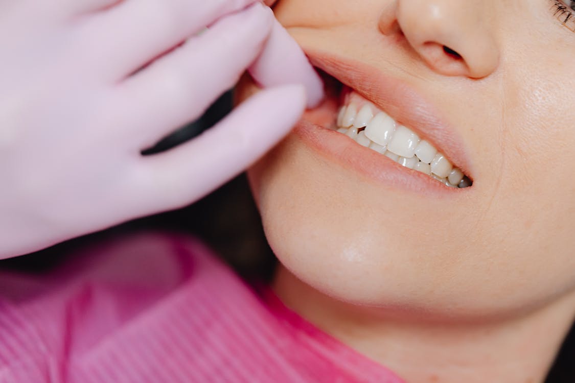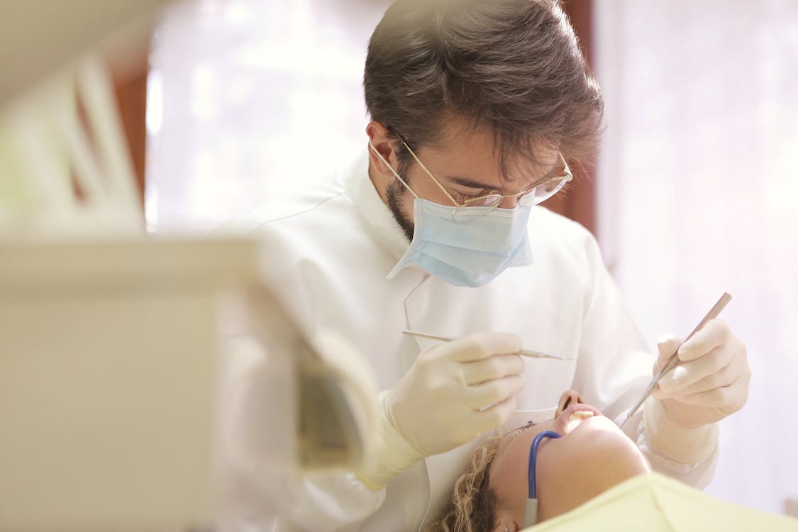
The temporomandibular joint — the hinge connecting your jawbone to your skull on each side of your face — is one of the most complex and frequently used joints in the human body. It opens and closes roughly 2,000 times per day during talking, chewing, yawning, and swallowing, combining rotational and sliding movements that no other joint in the body replicates. When this joint malfunctions, the resulting condition — broadly called temporomandibular joint disorder or TMJ disorder — can produce pain, clicking, locking, and functional limitations that affect everything from eating to speaking to sleeping.
More than 10 million Americans experience TMJ disorders at any given time, with women affected roughly twice as often as men. The condition ranges from mild, self-resolving episodes of jaw discomfort to chronic, debilitating pain that significantly impairs quality of life. Understanding the anatomy, causes, and available treatments helps those affected navigate a condition that is frequently misdiagnosed, overtreated, or undertreated depending on which provider they consult.
Understanding the TMJ
The temporomandibular joint is a bilateral joint — one on each side of the face, just in front of each ear. Each joint consists of the mandibular condyle (the rounded top of the jawbone), the temporal bone of the skull (which forms the socket), and an articular disc made of fibrocartilage that sits between the two bone surfaces, cushioning and guiding movement.
This disc is critical to normal joint function. During mouth opening, the disc slides forward along with the condyle, preventing bone-on-bone contact and distributing mechanical forces across the joint surfaces. When the disc displaces, degenerates, or fails to track properly with the condyle, the characteristic symptoms of TMJ disorder develop.
Surrounding muscles — including the masseter, temporalis, medial pterygoid, and lateral pterygoid — power jaw movement and stabilize the joint during function. These muscles generate tremendous force during chewing, with the masseter muscle producing up to 200 pounds of force on the back molars. Dysfunction in these muscles, whether from overuse, tension, or injury, can produce pain that mimics or accompanies joint problems.
Types of TMJ Disorders
TMJ disorders fall into three overlapping categories, and many patients experience elements of more than one type simultaneously.
Myofascial Pain
The most common form of TMJ disorder involves pain originating in the muscles that control jaw movement. Myofascial pain can produce aching in the jaw, face, temple, and ear areas, headaches that mimic tension-type headaches, pain that worsens with chewing or wide mouth opening, and referred pain to areas that seem unconnected to the jaw. This muscular form of TMJ disorder frequently develops from habitual clenching, grinding, or tension-holding patterns in the jaw muscles, often without the patient's awareness.
Internal Derangement
Internal derangement refers to problems with the articular disc — typically displacement from its normal position between the condyle and temporal bone. A displaced disc may produce clicking or popping sounds when the mouth opens and closes, as the condyle moves on and off the disc. If the disc displaces completely and the condyle cannot recapture it during opening, the jaw may lock in either an open or closed position.
Disc displacement with reduction (the disc pops back into place during opening, producing a click) is common and often painless. Disc displacement without reduction (the disc stays displaced, potentially limiting opening) is more problematic and may require treatment.
Degenerative Joint Disease
Arthritis affecting the TMJ — whether osteoarthritis from wear and tear or inflammatory arthritis from conditions like rheumatoid arthritis — damages the joint surfaces and can produce pain, crepitus (grinding or grating sounds), stiffness, and progressive limitation of jaw movement. This form of TMJ disorder is more common in older adults and those with systemic inflammatory conditions.
Common Causes and Risk Factors
TMJ disorders rarely have a single identifiable cause. Instead, multiple factors typically converge to produce symptoms.
Bruxism — grinding or clenching the teeth — ranks among the most significant contributing factors. Daytime clenching, often unconscious and stress-driven, subjects the jaw muscles and joint to sustained excessive force. Nighttime grinding generates even greater forces over prolonged periods without the protective feedback that consciousness provides. The National Institute of Dental and Craniofacial Research identifies bruxism as a primary contributor to TMJ symptoms.
Physical trauma to the jaw — from sports injuries, motor vehicle accidents, dental procedures, or falls — can damage the joint, disc, or surrounding structures. Whiplash injuries that do not directly involve the jaw can still strain the TMJ through rapid forced jaw movement during the head's acceleration and deceleration.
Stress and psychological factors play an underappreciated role. Emotional stress promotes muscle tension throughout the body, with the jaw muscles being a common site of tension-holding. Anxiety, depression, and poor stress management correlate significantly with TMJ disorder severity and treatment outcomes.
Occlusal factors — how the upper and lower teeth fit together — have been historically overemphasized as a cause of TMJ disorders. While severely misaligned bites can contribute to joint strain, the relationship between bite alignment and TMJ symptoms is less direct than previously believed. Irreversible dental treatments aimed at correcting bite alignment to treat TMJ disorders are no longer recommended as first-line interventions.
Hormonal factors likely contribute to the female predominance of TMJ disorders. Estrogen receptors have been identified in the TMJ, and symptom fluctuation correlating with menstrual cycles has been documented in some patients. Oral contraceptive use and hormone replacement therapy have been associated with increased TMJ symptom prevalence in some studies.
Connective tissue disorders including Ehlers-Danlos syndrome and other hypermobility conditions increase TMJ disorder risk by creating joint laxity that allows excessive movement and disc displacement.
Symptoms That Signal TMJ Dysfunction
The symptom profile of TMJ disorders extends well beyond jaw pain, which contributes to the frequency of misdiagnosis.
Primary symptoms include pain or tenderness in the jaw, face, or in and around the ear during chewing, speaking, or wide mouth opening. Clicking, popping, or grating sounds in the joint when moving the jaw are common, though painless clicking alone without other symptoms may not require treatment. Difficulty or discomfort while chewing, a sensation that the upper and lower teeth do not fit together properly, and locking of the joint that makes it difficult to open or close the mouth all indicate TMJ involvement.
Secondary symptoms that patients may not associate with a jaw problem include tension-type headaches, particularly upon waking. Ear symptoms including fullness, pain, ringing (tinnitus), and even dizziness can originate from TMJ dysfunction because of the joint's proximity to ear structures. Neck and shoulder pain frequently accompanies TMJ disorders through interconnected muscle chains. Tooth pain without dental pathology sometimes represents referred pain from TMJ muscular dysfunction.
Diagnosis
Accurate diagnosis requires differentiating TMJ disorders from the many conditions that produce similar symptoms — ear infections, sinus problems, dental pathology, trigeminal neuralgia, and tension headaches can all mimic TMJ symptoms.
A clinical examination evaluates jaw range of motion, joint sounds, muscle tenderness, and pain reproduction with specific movements. The American Dental Association supports a conservative diagnostic approach that begins with clinical history and physical examination before advancing to imaging.
Imaging studies are reserved for cases where clinical findings suggest structural abnormalities. Panoramic X-rays provide an overview of the jaw and teeth. Cone-beam CT scanning offers detailed three-dimensional views of the bony joint structures. MRI is the gold standard for visualizing soft tissue components, particularly the articular disc, and is used when disc displacement or other soft tissue pathology is suspected.
Home Care and Self-Management
The majority of TMJ disorders respond to conservative management, and professional guidelines strongly recommend exhausting non-invasive approaches before considering more aggressive treatments.
Jaw Exercises
Targeted exercises can reduce pain, improve range of motion, and strengthen the muscles that stabilize the TMJ. These exercises should be performed gently and stopped if they increase pain.
Relaxed jaw position: Rest the tongue lightly on the roof of the mouth behind the upper front teeth. Allow the teeth to separate and the jaw muscles to relax. This position, practiced throughout the day whenever jaw tension is noticed, retrains the muscles away from clenching habits.
Goldfish exercises (partial opening): Place one finger on the TMJ in front of each ear and one finger on the chin. Drop the lower jaw halfway open and then close. Practice this movement six times in one set, performing six sets daily. The fingers on the TMJ provide proprioceptive feedback that helps maintain smooth, centered jaw movement.
Goldfish exercises (full opening): Same positioning as partial opening, but allow the jaw to open fully. Perform six repetitions per set, six sets daily. If full opening produces clicking or catching, return to partial opening until smooth motion is achieved.
Chin tucks: Pull the chin straight back, creating a double chin appearance, while keeping the shoulders back and chest up. Hold for three seconds, repeat ten times. This exercise addresses the forward head posture that commonly accompanies TMJ disorders and contributes to muscle tension.
Resisted opening: Place a thumb under the chin. Open the mouth slowly while applying gentle resistance with the thumb. Hold for three to six seconds, then close. This isometric exercise strengthens the muscles responsible for controlled jaw opening.
Resisted closing: Place fingers on the front of the lower teeth. Close the mouth while applying gentle downward resistance with the fingers. This strengthens the muscles used for biting and chewing in a controlled manner.
Side-to-side movement: Place a quarter-inch object (like a stack of tongue depressors) between the front teeth. Slowly move the jaw from side to side. As the exercise becomes easier, increase the thickness of the object. This exercise improves lateral jaw mobility and strengthens the lateral pterygoid muscles.
Heat and Cold Therapy
Applying moist heat (warm towel or heating pad) to the jaw muscles for 15 to 20 minutes relaxes tense muscles and improves blood flow. Ice packs wrapped in a cloth and applied for 10 to 15 minutes reduce acute inflammation and numb pain. Alternating between heat and cold — 10 minutes of heat followed by 5 minutes of cold — can be particularly effective for combined muscular and inflammatory symptoms.
Dietary Modifications
Temporarily shifting to softer foods reduces the mechanical demands on the jaw during acute pain episodes. Cutting food into small pieces, avoiding chewy or hard items, and minimizing wide mouth opening during eating allow the joint and muscles to rest while healing occurs. This is a temporary strategy during flare-ups rather than a permanent dietary change.
Stress Management
Because stress-driven muscle tension is a primary driver of myofascial TMJ pain, stress management directly impacts symptoms. Progressive muscle relaxation, meditation, biofeedback, cognitive behavioral therapy, and regular exercise all reduce the baseline tension level in jaw muscles. Developing awareness of daytime clenching habits — checking jaw tension periodically throughout the day and consciously releasing — interrupts the sustained muscle contraction that produces pain.
Professional Treatment Options
Oral Appliances
Custom-fitted occlusal splints, commonly called night guards or bite guards, protect teeth from grinding damage and may reduce muscle activity during sleep. Stabilization splints that cover all the teeth on one arch are the most commonly prescribed and best-studied appliance type. These devices should be fabricated by a dental professional to ensure proper fit — ill-fitting over-the-counter guards can actually worsen TMJ symptoms by altering bite forces unpredictably.
Physical Therapy
Physical therapists specializing in orofacial pain provide manual therapy techniques including joint mobilization, myofascial release, and trigger point therapy that can reduce pain and improve jaw mobility. Modalities including ultrasound, electrical stimulation, and dry needling offer additional tools for managing muscular symptoms. A physical therapy program typically includes supervised treatment sessions combined with a home exercise program.
Medications
Over-the-counter pain relievers including ibuprofen and naproxen provide anti-inflammatory and analgesic effects for acute flare-ups. Muscle relaxants prescribed for short-term use can break the cycle of sustained muscle contraction. Low-dose tricyclic antidepressants, used at doses below the antidepressant range, have demonstrated effectiveness for chronic TMJ pain through their effects on pain processing pathways.
Botulinum toxin injections into the masseter and temporalis muscles have shown promise for TMJ disorders driven by muscle hyperactivity and bruxism. By temporarily weakening the muscles responsible for clenching, these injections can reduce pain and decrease the forces applied to the joint. Effects typically last three to four months before repeat treatment is needed.
Arthrocentesis
This minimally invasive procedure involves inserting needles into the joint space and irrigating with sterile fluid to wash out inflammatory debris and break up adhesions that may be limiting joint movement. Arthrocentesis is performed under local anesthesia and has a favorable risk-benefit profile for patients who have not responded to conservative treatment.
Surgery
Surgical intervention is reserved for the small minority of patients with structural abnormalities that do not respond to conservative management. Open joint surgery, arthroscopic surgery, and total joint replacement are available for severe cases but carry risks including nerve damage, scarring, and the possibility of incomplete symptom relief. Professional guidelines strongly recommend that irreversible surgical procedures be considered only after thorough conservative treatment trials have failed.
When to Seek Professional Help
While many TMJ symptoms resolve with self-care, certain situations warrant professional evaluation. Persistent pain lasting more than two weeks despite home management, progressive limitation of jaw opening, a jaw that locks in open or closed position, new onset of hearing changes or dizziness accompanying jaw symptoms, and pain that interferes with eating, speaking, or sleeping should all prompt evaluation by a dentist or physician experienced in TMJ disorders.
TMJ disorders can be frustrating precisely because they inhabit the space between dental and medical care, and patients sometimes bounce between providers without finding one who takes ownership of the diagnosis and treatment. Seeking a provider specifically experienced in temporomandibular disorders — whether a general dentist with TMJ expertise, an orofacial pain specialist, or a maxillofacial surgeon — ensures that evaluation and treatment are guided by current evidence rather than outdated approaches that may cause more harm than benefit.
Sources and Further Reading
Health and Beyond uses reputable medical and scientific sources where possible. These links support or expand on the topics discussed above.